Diagram Of Liver Gallbladder And Pancreas / Thin walled muscular sac on ventral surface of liver.. Thin walled muscular sac on ventral surface of liver. The stomach, gallbladder, and pancreas are three of the most important digestive organs in the human body. In vertebrates, the gallbladder is a small hollow organ where bile is stored and concentrated before it is released into the small intestine. The pancreas, gallbladder, and liver are identified as accessory organs. Liver, gallbladder, and pancreas by dr.
Liver, pancreas doctors and health care workers across many disciplines. The understanding of liver anatomy enables a surgeon to accurately locate and safely remove suspected liver tumours. The gallbladder, a saclike structure appended to the liver, concentrates and stores bile for use by the small intestine in fat digestion. The pancreas, gallbladder, and liver are identified as accessory organs. Several peritoneal ligaments support the position of the liver:
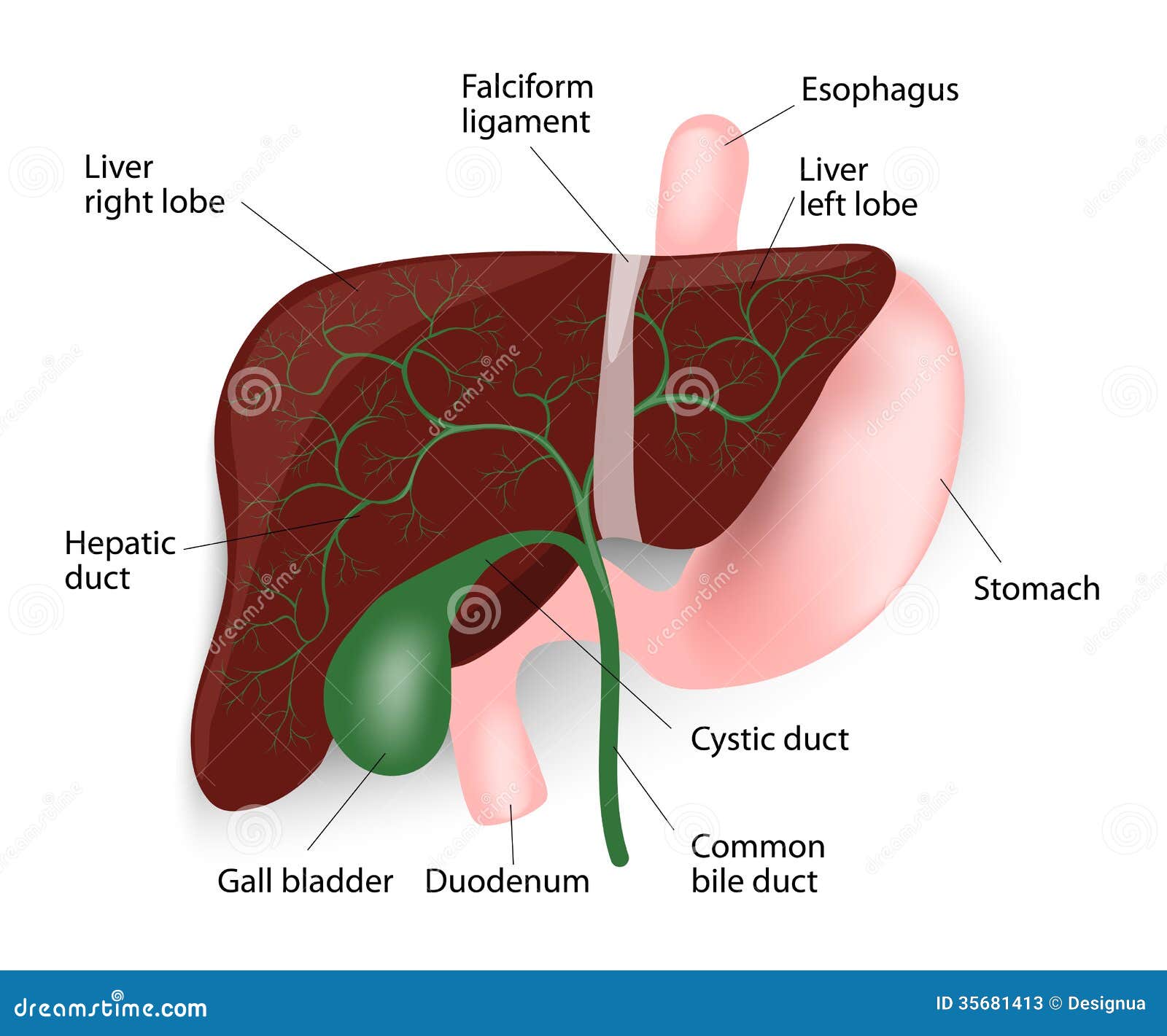
Liver Gallbladder Esophagus Stomach And Duodenu Stock Vector Illustration Of Anatomy Bile 35681413 from thumbs.dreamstime.com Liver, gallbladder and pancreas pp. They are secretory glands that are associated with the alimentary system. Liver, pancreas doctors and health care workers across many disciplines. It is joined by the common hepatic duct coming from the liver and cystic duct coming from the gallbladder. The aim of the abc of liver, pancreas and gall bladder is to provide an overview of these diseases. The bile is manufacturing continuously in the liver travels the bile forced to out to the duodenum via common bile duct and pancreatic duct. The duodenum or the first part of the small intestine receives a mixture. Thin walled muscular sac on ventral surface of liver.
Want to learn more about it?
Want to learn more about it? Advertisements help pay for this website. Branch through ct and empty into the sinusoids (o2 rich. Discuss the cellular structure of an exocrine pancreatic acinus and its function. This is a diagram showing the relationship between the liver, gallbladder, bile duct the pancreas and the pancreatic duct (the tube that drains the pancreatic juices) are in very. Liver, pancreas doctors and health care workers across many disciplines. Thin walled muscular sac on ventral surface of liver. How does the duodenum respond to the pre They are secretory glands that are associated with the alimentary system. Bacteria, fungi, and parasites also may cause hepatitis. A number of disorders can occur in the biliary system. Discriminate between exocrine and endocrine pancreas. Composition of gallbladder bile composition of gallbladder bile.
In vertebrates, the gallbladder is a small hollow organ where bile is stored and concentrated before it is released into the small intestine. Liver, gallbladder, and pancreas by dr. The liver is divided into 8 segments based on its blood supply. The function of the gallbladder is to store the dilute bile it receives from the hepatic duct, concentrate it. Functions to store and concentrate bile by absorbing water and ions.
Illustration Of The Normal Development Of The Pancreas A B Dorsal Download Scientific Diagram from www.researchgate.net What are the four functional groups of liver components? This is a diagram showing the relationship between the liver, gallbladder, bile duct the pancreas and the pancreatic duct (the tube that drains the pancreatic juices) are in very. Branch through ct and empty into the sinusoids (o2 rich. Draw a diagram depicting human alimentary canal and label on it. 5 circulation to and from liver where does blood inferior side of liver at the porta hepatis where do the portal vein and hepatic artery empty? Chapter 5 liver, gallbladder, and pancreas the liver maintains the physiologic and metabolic balance of the body. Discuss the cellular structure of an exocrine pancreatic acinus and its function. The head of the pancreas lies in the duodenal loop and the tail extends across the abdominal cavity to the spleen.
Discuss the cellular structure of an exocrine pancreatic acinus and its function.
The pancreas, gallbladder, and liver are identified as accessory organs. Advertisements help pay for this website. This means they play a role in digestion, but they're separate from the digestive tract. Bacteria, fungi, and parasites also may cause hepatitis. The understanding of liver anatomy enables a surgeon to accurately locate and safely remove suspected liver tumours. Accessory duct of the pancreas. Study heather fahey's gi, liver, gallbladder, pancreas flashcards now! The bile ducts link the liver to the gallbladder as well, so that some of the bile which is made can be stored for future use. The gallbladder, a saclike structure appended to the liver, concentrates and stores bile for use by the small intestine in fat digestion. Some of the most common are highlighted below. Liver gallbladder and pancreas model. It travels posteriorly, through the head of the pancreas to merge with the pancreatic duct and then onto the superior aspect of the duodenum to form the hepatopancreatic ampulla. Liver, gallbladder and pancreas accessory organs associated with small intestine liver digestive function is production of bile bile fat emulsifier gallbladder.
Pancreas, liver, and gallbladder | chapter 15. Some of the most common are highlighted below. Branch through ct and empty into the sinusoids (o2 rich. 5 circulation to and from liver where does blood inferior side of liver at the porta hepatis where do the portal vein and hepatic artery empty? Advertisements help pay for this website.
File Blausen 0428 Gallbladder Liver Pancreas Location Png Wikimedia Commons from upload.wikimedia.org In vertebrates, the gallbladder is a small hollow organ where bile is stored and concentrated before it is released into the small intestine. The duodenum or the first part of the small intestine receives a mixture. To this end it contains helpful algorithms for diagnosing and treating common diseases. Sharply localized or a diffuse narrowing. Pancreas, liver, and gallbladder | chapter 15. Liver, gallbladder and pancreas accessory organs associated with small intestine liver digestive function is production of bile bile fat emulsifier gallbladder. Liver the histological organization of the liver is complex. What are the four functional groups of liver components?
Liver, pancreas doctors and health care workers across many disciplines.
It is joined by the common hepatic duct coming from the liver and cystic duct coming from the gallbladder. Thin walled muscular sac on ventral surface of liver. The bile ducts link the liver to the gallbladder as well, so that some of the bile which is made can be stored for future use. To know the histology of the liver, including the hepatic lobule, hepatic acinus, hepatic parenchyma, portal area, and histological features of the vascular and biliary systems. How does the duodenum respond to the pre Functions to store and concentrate bile by absorbing water and ions. The stomach, gallbladder, and pancreas are three of the most important digestive organs in the human body. More prominent following contraction after a fatty meal. The biliary system, including the liver, pancreas and gallbladder, form a part of the body's digestive system that is responsible for nutrient absorption and waste disposal. The bile is manufacturing continuously in the liver travels the bile forced to out to the duodenum via common bile duct and pancreatic duct. This is a diagram showing the relationship between the liver, gallbladder, bile duct the pancreas and the pancreatic duct (the tube that drains the pancreatic juices) are in very. Right and left kidneys (silhouette). Several peritoneal ligaments support the position of the liver:
The head of the pancreas lies in the duodenal loop and the tail extends across the abdominal cavity to the spleen diagram of liver. They are secretory glands that are associated with the alimentary system.


Post a Comment